微信扫码


 上海宇劲生物孙经理


·产品描述
最近证明5-甲酰胞嘧啶(5-fc)存在于哺乳动物的组织和细胞内,现已被定义为第七种碱基,来源于5-羟甲基胞嘧啶的氧化;已被证明参与DNA去甲基化的过程.虽然5-fc的具体功能还不清楚,但是在小鼠胚胎植入发育过程中已经证明具有促进复制的功能.目前5-fc已被作为DNA去甲基化的活性标志.
MethylFlash 5-甲酰胞嘧啶DNA定量试剂盒是包含实验所需试剂,基于比色检测原理来定量样本5-fc的一套完整组件.适用于各种样本分离的DNA.实验过程中DNA被绑定于孔板内,通过捕获抗体和检测抗体来比色定量样本中5-fc的含量.检测波长为450nm.
·产品特点
1、快速简单的操作步骤,完成实验不超过3小时45分钟;
2、高灵敏度,其检测极限为1pg;
3、高特异性,只能识别5-fc;
4、适用于细胞或者组织分离DNA样本,无需消化和水解;
5、包含阴阳性对照,适用于各种种属;
6 适用于高通量试验;
7、可重复性高.
The MethylFlash™ 5-Formylcytosine (5-fC) DNA Quantification Kit (Colorimetric) is a complete set of optimized buffers and reagents to colorimetrically quantify 5-formylcytosine (5-fC) in a microplate-based format. It is suitable for use with DNA isolated from any species (mammals, plants, fungi, bacteria, viruses, etc.) in a variety of forms including, but not limited to, cultured cells, fresh and frozen tissues, paraffin-embedded tissues, and body fluid samples. The kit has the following advantages and features:
Background Information
5-formylcytosine (5-fC) has recently been found in mammalian tissue and cells and has been classified as a seventh DNA base. It is formed by the oxidation of 5-hydroxymethylcytosine through TET hydroxylases and has been demonstrated to be an intermediate of the DNA demethylation process. The function of 5-fC in gene regulation is not yet entirely clear, but it has been shown that 5-fC exhibits replication-dependent dilution during mouse pre-implantation development and could be functional in regulating pre-implantation development as a whole. The detection of 5-fC in various tissues and cells is important because 5-fC could be a marker to indicate active DNA demethylation. 5-fC can also be directly excised by Thymine DNA glycosylase (TDG) to allow subsequent base excision repair (BER) processing which converts modified cytosine back to its unmodified state.
Several chromatography-based techniques such as HPLC-MS are used for the detection of 5-fC in tissue and cells. However, these methods are time consuming, require large amounts of DNA and have low throughput with high costs. To address these problems, Epigentek offers the MethylFlash™ 5-Formylcytosine (5-fC) DNA Quantification Kit (Colorimetric), which uses a unique microplate-based procedure to directly quantify 5-fC.
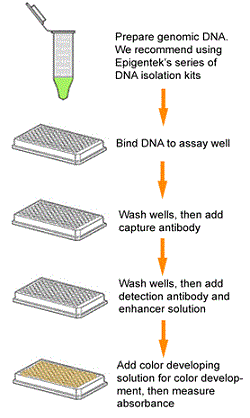
Principle & Procedure
The MethylFlash™ 5-Formylcytosine (5-fC) DNA Quantification Kit (Colorimetric) contains all the reagents necessary for the quantification of 5-fC. In this assay, DNA is bound to strip-wells that are specifically treated to have a high DNA affinity. 5-fC is detected using capture and detection antibodies. The detected signal is enhanced and then quantified colorimetrically by reading the absorbance in a microplate spectrophotometer at a wavelength of 450 nm. The amount of 5-fC is proportional to the OD intensity measured.
Starting Materials & Input Amount
The detection limit of the input DNA can be as low as 1 pg of 5-fC with a dynamic range from 10 pg to 400 pg (see Fig. 2). There is no cross-reactivity to cytosine, 5-mC, or 5-hmC within the indicated concentration range of the sample DNA (see Fig. 3).
Safe and Convenient
All the necessary reagents, including negative and positive controls, for the quantification of 5-fC are conveniently packaged in the kit. The direct colorimetric quantification of DNA samples in a 96-well microplate format eliminates the need for DNA digestion/denaturation, radioactivity, extraction, or chromatography.
Easy, Fast, and Flexible
The entire colorimetric assay has easy-to-follow steps for convenience and speed, allowing it to be completed in just 3 hours and 45 minutes. The strip-well microplate format allows for a flexible assay for manual or high throughput analysis. Universal positive and negative controls are included with the kit for the quantification of 5-fC from any species, such as mammals, plants, fungi, bacteria, and viruses.
Responsive, Reliable, and Practical
Based on its working principle and the microplate format, the kit can be practically and routinely used for any species and for a variety of forms including cultured cells, fresh and frozen tissues, and paraffin-embedded tissues. To demonstrate the capabilities of the kit, it has been successfully used for quantifying the content of 5-fC in DNA from human brain tissue, mouse brain tissue, rectal tissue, and rectal cancer. The percentage of 5-fC measured by the kit is parallel and comparable to that detected by LC-MS/MS methods (see Fig. 4).
Product Citations
Burghardt KJ et. al. (October 2019). Skeletal muscle DNA methylation modifications and psychopharmacologic treatment in bipolar disorder. Eur Neuropsychopharmacol.
Chen D et. al. (August 2019). Polymerization retardation isothermal amplification (PRIA): a strategy enables sensitively quantify genome-wide 5-methylcytosine oxides rapidly on handy instruments with nanoscale sample input. Nucleic Acids Res.
Tardu M et. al. (October 2018). Identification and quantification of modified nucleosides in Saccharomyces cerevisiae mRNAs bioRxiv.
M Katayama et. al. (July 2018). Determination of 5-Metylcytosine, 5-Hydroxymethylcytosine, and 5-Formylcytosine Expression in Mobile and Immobile Sperm From One Ejaculation: Analysis of Relationsips With Sperm Properties using Partial Least Square Regression Indian Journal of Research. 7(4)
Swathy B et. al. (January 2018). Understanding the influence of antipsychotic drugs on global methylation events and its relevance in treatment response. Epigenomics.
Çelik SU et. al. (April 2016). The sensitivity of 5-formylcytosine to doxorubicin regardless of DNA damage Turk J Biol. 40
Neri F et. al. (February 2015). Single-Base Resolution Analysis of 5-Formyl and 5-Carboxyl Cytosine Reveals Promoter DNA Methylation Dynamics. Cell Rep.
Chowdhury B et. al. (December 2014). Quantification of 5-methylcytosine, 5-hydroxymethylcytosine and 5-carboxylcytosine from the blood of cancer patients by an enzyme-based immunoassay. Anal Chim Acta. 852:212-7.
Kang KA et. al. (April 2014). Epigenetic modification of Nrf2 in 5-fluorouracil-resistant colon cancer cells: involvement of TET-dependent DNA demethylation. Cell Death Dis. 5:e1183.
Dhliwayo N et. al. (April 2014). Parp inhibition prevents ten eleven translocase enzyme activation and hyperglycemia induced DNA demethylation. Diabetes.
 购物车
购物车