微信扫码


 上海宇劲生物孙经理


| 品牌 | 货号 | 产品名称 | 规格 |
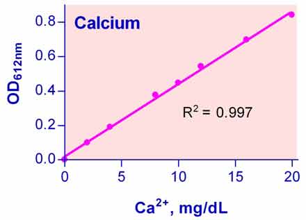
| BioAssay Systems | DICA-500 | QuantiChrom™ Calcium Assay Kit 钙离子测试盒 | 500T |
说明书:
·相关文献
Cruz, A.C.C. et al (2019). Retinoic acid increases the effect of bone morphogenetic protein type 2 on osteogenic differentiation of human adipose-derived stem cells. Journal of Applied Oral Science 27:e20180317. Assay: Calcium in human mesenchymal stem cells.
Li, Y., et al. (2019). Biofunctionalization of decellularized porcine aortic valve with OPG-loaded PCL nanoparticles for anti-calcification. RSC advances, 9(21), 11882-11893. Assay: Calcium in rats bone marrow.
Schanstra, J. P.et al. (2019). Systems biology identifies cytosolic PLA2 as a target in vascular calcification treatment. JCI insight, 4(10) pii: 125638. Assay: Calcium in human plasma.
Schelski, N., Luong, T. T., Lang, F., Pieske, B., Voelkl, J., & Alesutan, I. (2019). SGK1-dependent stimulation of vascular smooth muscle cell osteo-/chondrogenic transdifferentiation by interleukin-18. Pflugers Archiv-European Journal of Physiology, 471(6):889-899. Assay: Calcium in human aortic smooth muscle cells.
Tsolaki, E., Didierlaurent, L., Muller, E., Rottmar, M., Latif, N., Chester, A. H. & Bertazzo, S. (2019). Electron microscopy characterization of minerals formed in vitro by human bone cells and vascular smooth muscle cells. BioRxiv, 661785. Assay: Calcium in human bone cells.
Bora, S. A., Kennett, M. J., Smith, P. B., Patterson, A. D., & Cantorna, M. T. (2018). The gut microbiota regulates endocrine vitamin D metabolism through fibroblast growth factor 23. Frontiers in immunology, 9, 408. Assay: Calcium in mice cells.
Bouyoucos, I. A., Talwar, B. S., Brooks, E. J., Brownscombe, J. W., Cooke, S. J., Suski, C. D., & Mandelman, J. W. (2018). Exercise intensity while hooked is associated with physiological status of longline-captured sharks. Conservation physiology, 6(1), coy074. Assay: Calcium in shark plasma calcium.
Gurel Pekozer, G., Ramazanoglu, M., Schlegel, K. A., Kok, F. N., & Torun Kose, G. (2018). Role of STRO-1 sorting of porcine dental germ stem cells in dental stem cell-mediated bone tissue engineering. Artificial cells, nanomedicine, and biotechnology, 46(3), 607-618. Assay: Calcium in porcine teeth.
Kagi, L., Bettoni, C., Pastor-Arroyo, E. M., Schnitzbauer, U., Hernando, N., & Wagner, C. A. (2018). Regulation of vitamin D metabolizing enzymes in murine renal and extrarenal tissues by dietary phosphate, FGF23, and 1, 25 (OH) 2D3. PloS one, 13(5), e0195427. Assay: Calcium in mice spot urine.
Ma, G. T., Lee, S. K., Park, K. K., Park, J., Son, S. H., Jung, M., & Chung, W. Y. (2018). Artemisinin-Daumone Hybrid Inhibits Cancer Cell-Mediated Osteolysis by Targeting Cancer Cells and Osteoclasts. Cellular Physiology and Biochemistry, 49(4), 1460-1475. Assay: Calcium in mice serum.
Onal, M., Carlson, A. H., Thostenson, J. D., Benkusky, N. A., Meyer, M. B., Lee, S. M., & Pike, J. W. (2018). A Novel Distal Enhancer Mediates Inflammation-, PTH-, and Early Onset Murine Kidney Disease-Induced Expression of the mouse Fgf23 Gene. JBMR plus, 2(1), 31-46. Assay: Calcium in mice maxillary blood.
Roh, J. Y., & Kim, K. R. (2018). Anti-inflammatory effect of new calcium hydroxide paste containing silicon-substituted hydroxyapatite in lipopolysaccharide-stimulated macrophages. J Korean Soc Dent Hyg 18(4), 423-432. Assay: Calcium in plant.
Townsend, J. M., Zabel, T. A., Feng, Y., Wang, J., Andrews, B. T., Nudo, R. J. & Detamore, M. S. (2018). Effects of tissue processing on bioactivity of cartilage matrix-based hydrogels encapsulating osteoconductive particles. Biomedical Materials, 13(3), 034108. Assay: Calcium in rats bone marrow.
Vollersen, N., Hermans-Borgmeyer, I., Cornils, K., Fehse, B., Rolvien, T., Triviai, I. & Yorgan, T. A. (2018). High Bone Turnover in Mice Carrying a Pathogenic Notch2 Mutation Causing Hajdu-Cheney Syndrome. Journal of Bone and Mineral Research, 33(1), 70-83. Assay: Calcium in mice serum.
Aimaiti, A., Maimaitiyiming, A., Boyong, X., Aji, K., Li, C., & Cui, L. (2017). Low-dose strontium stimulates osteogenesis but high-dose doses cause apoptosis in human adipose-derived stem cells via regulation of the ERK1/2 signaling pathway. Stem cell research & therapy, 8(1), 282. Assay: Calcium in human bone marrow.
BARINDA, A. J., Ikeda, K., Hirata, K. I., & Emoto, N. (2017). Macrophages Highly Express Carbonic Anhydrase 2 and Play a Significant Role in Demineralization of the Ectopic Calcification. Kobe Journal of Medical Sciences, 63(2), E45. Assay: Calcium in culture plate fibronectin.
Go, Y. Y., Kim, S. E., Cho, G. J., Chae, S. W., & Song, J. J. (2017). Differential effects of amnion and chorion membrane extracts on osteoblast-like cells due to the different growth factor composition of the extracts. PloS one, 12(8), e0182716. Assay: Calcium in human amniotic membrane.
Hu, K., Sun, H., Gui, B., & Sui, C. (2017). Gremlin-1 suppression increases BMP-2-induced osteogenesis of human mesenchymal stem cells. Molecular medicine reports, 15(4), 2186-2194. Assay: Calcium in human bone marrow.
Kim, E. C., Park, J., Kwon, I. K., Lee, S. W., Park, S. J., & Ahn, S. J. (2017). Static magnetic fields promote osteoblastic/cementoblastic differentiation in osteoblasts, cementoblasts, and periodontal ligament cells. Journal of periodontal & implant science, 47(5), 273-291. Assay: Calcium in human fetal osteoblast.
Paul, S., Gangwar, A., Bhargava, K., & Ahmad, Y. (2017). Deciphering Molecular Cascades In A Novel Acclimatization Strategy For Rapid Ascent To High Altitude. bioRxiv, 145342. Assay: Calcium in Sprague Dewley rats lung tissue/plasma.
Vanacker, N., Ollier, S., Beaudoin, F., Blouin, R., & Lacasse, P. (2017). Effect of inhibiting the lactogenic signal at calving on milk production and metabolic and immune perturbations in dairy cows. Journal of dairy science, 100(7), 5782-5791. Assay: Calcium in Holstein Cows serum.
Wang, F., Johnson, R. L., DeSmet, M. L., Snyder, P. W., Fairfax, K. C., & Fleet, J. C. (2017). Vitamin D Receptor-Dependent Signaling Protects Mice From Dextran Sulfate Sodium-Induced Colitis. Endocrinology, 158(6), 1951-1963. Assay: Calcium in mice intestinal cells.
Birgani, Z. T., van Blitterswijk, C. A., & Habibovic, P. (2016). Monolithic calcium phosphate/poly (lactic acid) composite versus calcium phosphate-coated poly (lactic acid) for support of osteogenic differentiation of human mesenchymal stromal cells. Journal of Materials Science: Materials in Medicine, 27(3), 54. Assay: Calcium in monolithic PLA/CaP composite.
Cai, T., Sun, D., Duan, Y., Wen, P., Dai, C., Yang, J., & He, W. (2016). WNT/beta-catenin signaling promotes VSMCs to osteogenic transdifferentiation and calcification through directly modulating Runx2 gene expression. Experimental cell research, 345(2), 206-217. Assay: Calcium in rats smooth muscles.
Corcoran, A., Bermudez, M. A., Seoane, S., Perez-Fernandez, R., Krupa, M., Pietraszek, A. & Marcinkowska, E. (2016). Biological evaluation of new vitamin D2 analogues. The Journal of steroid biochemistry and molecular biology, 164, 66-71. Assay: Calcium in mice serum.
Go, Y. Y., Kim, S. E., Cho, G. J., Chae, S. W., & Song, J. J. (2016). Promotion of osteogenic differentiation by amnion/chorion membrane extracts. Journal of applied biomaterials & functional materials, 14(2), 171-180. Assay: Calcium in human amniotic membrane.
Kim, W., et al. (2016). Calcium-sensing receptor promotes breast cancer by stimulating intracrine actions of parathyroid hormone-related protein. Cancer research, 76(18), 5348-5360. Assay: Calcium in mice serum.
Kose, S et al (2016). Evaluation of biocompatibility of random or aligned electrospun polyhydroxybutyrate scaffolds combined with human mesenchymal stem cells. Turkish Journal of Biology, 40(2), 410-419. Assay: Calcium in human myscenchymal cells.
Mao, J., Shi, X., Wu, Y. B., & Gong, S. Q. (2016). Identification of specific hydroxyapatite {001} binding heptapeptide by phage display and its nucleation effect. Materials, 9(8), 700. Assay: Calcium in human teeth.
Onal, M., St. John, H. C., Danielson, A. L., & Pike, J. W. (2016). Deletion of the distal Tnfsf11 RL-D2 enhancer that contributes to PTH-mediated RANKL expression in osteoblast lineage cells results in a high bone mass phenotype in mice. Journal of Bone and Mineral Research, 31(2), 416-429. Assay: Calcium in mice osteoblast cells.
Villa-Bellosta, R., Gonzalez-Parra, E., & Egido, J. (2016). Alkalosis and dialytic clearance of phosphate increases phosphatase activity: a hidden consequence of hemodialysis. PloS one, 11(7), e0159858. Assay: Calcium in human plasma.
Villa-Bellosta, R., Hamczyk, M. R., & Andres, V. (2016). Alternatively activated macrophages exhibit an anticalcifying activity dependent on extracellular ATP/pyrophosphate metabolism. American Journal of Physiology-Cell Physiology, 310(10), C788-C799. Assay: Calcium in mice smooth muscles.
Ballester-Lozano GF et al. (2015). Comprehensive biometric, biochemical and histopathological assessment of nutrient deficiencies in gilthead sea bream fed semi-purified diets. Br J Nutr. 114(5):713-26. Assay: calcium in fish plasma.
Chaumet-Riffaud P, et al (2010). Synthesis and application of lactosylated, 99mTc chelating albumin for measurement of liver function. Bioconjug Chem. 21(4):589-96. Assay: Calcium in mice liver tissue.
Jung GY, et al (2010). Effects of HA released calcium ion on osteoblast differentiation. J Mater Sci Mater Med. 21(5):1649-54. Assay: Calcium in mouse 3T3 cell.
Ou KL, et al (2010). Effects of the nanostructure and nanoporosity on bioactive nanohydroxyapatite/reconstituted collagen by electrodeposition. J Biomed Mater Res A. 92(3):906-12. Assay: Calcium in human stem cell.
Ponda MP,et al (2010). Moderate kidney disease inhibits atherosclerosis regression. Atherosclerosis.210(1):57-62. Assay: Calcium in mice serum.
Zarjou A, et al (2010). Ferritin ferroxidase activity: a potent inhibitor of osteogenesis. J Bone Miner Res. 25(1):164-72. Assay: Calcium in human osteoblasts cell.
Brand A, et al (2009). Calcium homeostasis is required for contact-dependent helical and sinusoidal tip growth in Candida albicans hyphae. Mol Microbiol. 71(5):1155-64. Assay: Calcium in cattle FBS.
Koreckij T, et al (2009). Dasatinib inhibits the growth of prostate cancer in bone and provides additional protection from osteolysis. Br J Cancer.101(2):263-8. Assay: Calcium in mice serum.
Villa-Bellosta R, Sorribas V (2009). Phosphonoformic acid prevents vascular smooth muscle cell calcification by inhibiting calcium-phosphate deposition. Arterioscler Thromb Vasc Biol. 29(5):761-6. Assay: Calcium in rat muscle cell.
Chanda, D et al (2008). Systemic osteoprotegerin gene therapy restores tumor-induced bone loss in a therapeutic model of breast cancer bone metastasis. Mol Ther. 16(5):871-8. Assay: Calcium in mouse serum.
He X, et al (2008). Effect of grafting RGD and BMP-2 protein-derived peptides to a hydrogel substrate on osteogenic differentiation of marrow stromal cells. Langmuir. 24(21):12508-16. Assay: Calcium in human marrow cell.
Henderson JA, et al (2008). Concurrent differentiation of marrow stromal cells to osteogenic and vasculogenic lineages. Macromol Biosci. 8(6):499-507. Assay: Calcium in human marrow cell.
Warotayanont R, et al (2008). Leucine-rich amelogenin peptide induces osteogenesis in mouse embryonic stem cells. Biochem Biophys Res Commun. 367(1):1-6. Assay: Calcium in mouse stem cell.
 购物车
购物车