微信扫码


 上海宇劲生物孙经理


| 品牌 | 货号 | 产品名称 | 规格 |
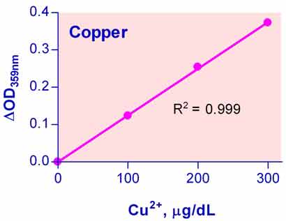
| BioAssay Systems | DICU-250 | QuantiChrom™ Copper Assay Kit 铜离子测试盒 | 250T |
说明书:
·相关文献
Parmar, A., Pascali, G., Lerra, L., Yee, E., Ahmed-Cox, A., Kimpton, K. & Liu, G. J. (2018). In vivo [64Cu] CuCl2 PET imaging reveals activity of Dextran-Catechin on tumor copper homeostasis. Theranostics, 8(20), 5645-5659. Assay: Copper in neuroblastoma cell.
Dikicioglu, D., & Oliver, S. G. (2017). Estimating global enzyme abundance levels from cofactor requirements: a model-based analysis of the iron metabolism in yeast. bioRxiv, 229104. Assay: Copper in S. cerevisiae cells.
Yee, E. M., Brandl, M. B., Pasquier, E., Cirillo, G., Kimpton, K., Kavallaris, M. & Vittorio, O. (2017). Dextran-Catechin inhibits angiogenesis by disrupting copper homeostasis in endothelial cells. Scientific reports, 7(1), 7638. Assay: Copper in human cells.
Vittorio, O., Brandl, M., Cirillo, G., Kimpton, K., Hinde, E., Gaus, K. & Haber, M. (2016). Dextran-Catechin: An anticancer chemically-modified natural compound targeting copper that attenuates neuroblastoma growth. Oncotarget 7(30): 47479-47493. Assay: Copper in human cells.
EL-Deeb, W. M., & El-Bahr, S. M. (2014). Selected Biochemical Indicators of Equine Rhabdomyolysis in Arabian Horses: Acute Phase Proteins and Trace Elements. Journal of Equine Veterinary Science 34(4): 484-488. Assay: Copper in horse serum.
Bartnikas TB (2012) Known and potential roles of transferrin in iron biology. Biometals 25(4):677-86. Assay: Copper in human protein.
Philips N et al (2012) Beneficial regulation of fibrillar collagens, heat shock protein-47, elastin fiber components, transforming growth factor-beta1, vascular endothelial growth factor and oxidative stress effects by copper in dermal fibroblasts. Connect Tissue Res. 53(5):373-8. Assay: Copper in human cell.
Piret JP et al (2012) Differential toxicity of copper (II) oxide nanoparticles of similar hydrodynamic diameter on human differentiated intestinal Caco-2 cell monolayers is correlated in part to copper release and shape. Nanotoxicology 6:789-803. Assay: Copper in human cell.
Piret, JP et al (2012). Copper (II) oxide nanoparticles penetrate into HepG2 cells, exert cytotoxicity via oxidative stress and induce pro-inflammatory response. Nanoscale 4(22): 7168-7184. Assay: Copper in human cell culture medium.
Lull ME, et al (2008). Plasma biomarkers in pediatric patients undergoing cardiopulmonary bypass. Pediatr Res. 63(6):638-44. Assay: Copper in human plasma.
 购物车
购物车