微信扫码


 上海宇劲生物孙经理


| 品牌 | 货号 | 产品名称 | 规格 |
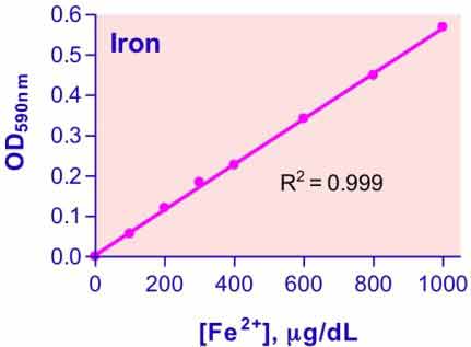
| BioAssay Systems | DIFE-250 | QuantiChrom™ Iron Assay Kit 铁离子测试盒 | 250T |
说明书:
·相关文献
Dichtl, S., Demetz, E., Haschka, D., Tymoszuk, P., Petzer, V., Nairz, M. & Theurl, I. (2019). Dopamine Is a Siderophore-Like Iron Chelator That Promotes Salmonella enterica Serovar Typhimurium Virulence in Mice. mBio, 10(1), e02624-18. Assay: Iron in mice serum.
Malhotra, H., Kumar, M., Chauhan, A. S., Dhiman, A., Chaudhary, S., Patidar, A. & Raje, M. (2019). Moonlighting Protein Glyceraldehyde-3-Phosphate Dehydrogenase: A Cellular Rapid-Response Molecule for Maintenance of Iron Homeostasis in Hypoxia. Cellular physiology and biochemistry: international journal of experimental cellular physiology, biochemistry, and pharmacology, 52(3), 517-531. Assay: Iron in mouse cells.
Cheli, V. T., Gonzalez, D. A. S., Marziali, L. N., Zamora, N. N., Guitart, M. E., Spreuer, V. & Paez, P. M. (2018). The Divalent Metal Transporter 1 (DMT1) is required for iron uptake and normal development of oligodendrocyte progenitor cells. Journal of Neuroscience, 38(43), 9142-9159. Assay: Iron in mice cells.
Efird, W. M., Fletcher, A. G., Draeger, R. W., Spang, J. T., Dahners, L. E., & Weinhold, P. S. (2018). Deferoxamine-Soaked Suture Improves Angiogenesis and Repair Potential After Acute Injury of the Chicken Achilles Tendon. Orthopaedic journal of sports medicine, 6(10), 2325967118802792. Assay: Iron in broiler chicks DFO.
Samba Mondonga, M., Calve, A., Mallette, F. A., & Santos, M. M. (2018). MyD88 regulates the expression of SMAD4 and the iron regulatory hormone hepcidin in hepatoma cells. Frontiers in cell and developmental biology, 6, 105. Assay: Iron in human kidney cells.
Worley, B. L., Kim, Y. S., Mardini, J., Zaman, R., Leon, K. E., Vallur, P. G. & Phaeton, R. (2018). GPx3 supports ovarian cancer progression by manipulating the extracellular redox environment. Redox Biology pii: S2213-2317(18)30891-7. Assay: Iron in human ovarian cells.
Asshoff, M., Petzer, V., Warr, M. R., Haschka, D., Tymoszuk, P., Demetz, E. & Fowles, P. (2017). Momelotinib inhibits ACVR1/ALK2, decreases hepcidin production, and ameliorates anemia of chronic disease in rodents. Blood, 129(13), 1823-1830. Assay: Iron in rat/mice serum.
Ma, X., Pham, V. T., Mori, H., MacDougald, O. A., Shah, Y. M., & Bodary, P. F. (2017). Iron elevation and adipose tissue remodeling in the epididymal depot of a mouse model of polygenic obesity. PloS one, 12(6), e0179889. Assay: Iron in mice serum.
Marks, E. S., Bonnemaison, M. L., Brusnahan, S. K., Zhang, W., Fan, W., Garrison, J. C., & Boesen, E. I. (2017). Renal iron accumulation occurs in lupus nephritis and iron chelation delays the onset of albuminuria. Scientific reports, 7(1), 12821. Assay: Iron in mice plasma.
Quan, Y. Y., Liu, Y. H., Lin, C. M., Wang, X. P., & Chen, T. S. (2017). Peroxynitrite dominates sodium nitroprusside-induced apoptosis in human hepatocellular carcinoma cells. Oncotarget 8(18): 29833-29845. Assay: Iron in cells.
Shang, Y. M., Wang, G. S., Sliney, D. H., Yang, C. H., & Lee, L. L. (2017). Light-emitting-diode induced retinal damage and its wavelength dependency in vivo. International journal of ophthalmology 10(2): 191-202. Assay: Iron in Sprague Dewley rats retina protein.
Sun, X., Zhao, Y., Jia, J., Xie, J., Cheng, J., Liu, H. & Fu, Y. (2017). Uninterrupted expression of CmSIT1 in a sclerotial parasite Coniothyrium minitans leads to reduced growth and enhanced antifungal ability. Frontiers in microbiology, 8, 2208. Assay: Iron in Coniothyrium minitans cells.
Szemraj, M., Oszajca, K., Szemraj, J., & Jurowski, P. (2017). MicroRNA expression analysis in serum of patients with congenital hemochromatosis and age-related macular degeneration (AMD). Medical science monitor: international medical journal of experimental and clinical research 23: 4050-4060. Assay: Iron in human serum.
Thongdee, P., & Na-Bangchang, K. (2017). The role of heme-oxygenase-1 in pathogenesis of cerebral malaria in the co-culture model of human brain microvascular endothelial cell and ITG Plasmodium falciparum-infected red blood cells. Asian Pacific journal of tropical medicine 10(1): 20-24. Assay: Iron in human brain cells.
Zhang, C. W., Tai, Y. K., Chai, B. H., Chew, K. C., Ang, E. T., Tsang, F. & Lim, K. L. (2017). Transgenic mice overexpressing the divalent metal transporter 1 exhibit iron accumulation and enhanced parkin expression in the brain. Neuromolecular medicine, 19(2-3), 375-386. Assay: Iron in mouse feces.
Hendricks, M. R., Lashua, L. P., Fischer, D. K., Flitter, B. A., Eichinger, K. M., Durbin, J. E. & Bomberger, J. M. (2016). Respiratory syncytial virus infection enhances Pseudomonas aeruginosa biofilm growth through dysregulation of nutritional immunity. Proceedings of the National Academy of Sciences, 113(6), 1642-1647. Assay: Iron in human cells.
Karoopongse, E., Marcondes, A. M., Yeung, C., Holman, Z., Kowdley, K. V., Campbell, J. S., & Deeg, H. J. (2016). Disruption of iron regulation after radiation and donor cell infusion. Biology of Blood and Marrow Transplantation, 22(7), 1173-1181. Assay: Iron in mouse serum.
Li, Y., Pan, K., Chen, L., Ning, J. L., Li, X., Yang, T. & Tao, G. (2016). Deferoxamine regulates neuroinflammation and iron homeostasis in a mouse model of postoperative cognitive dysfunction. Journal of neuroinflammation, 13(1), 268. Assay: Iron in mice cells.
Noguchi-Sasaki, M., Sasaki, Y., Shimonaka, Y., Mori, K., & Fujimoto-Ouchi, K. (2016). Treatment with anti-IL-6 receptor antibody prevented increase in serum hepcidin levels and improved anemia in mice inoculated with IL-6-producing lung carcinoma cells. BMC Cancer 16: 270. Assay: Iron in mouse serum.
Tamayo, E., Benabdellah, K., & Ferrol, N. (2016). Characterization of three new glutaredoxin genes in the arbuscular mycorrhizal fungus Rhizophagus irregularis: putative role of RiGRX4 and RiGRX5 in iron homeostasis. PloS one, 11(2), e0149606. Assay: Iron in yeast cells.
Theurl, I., Hilgendorf, I., Nairz, M., Tymoszuk, P., Haschka, D., Asshoff, M. & Sopper, S. (2016). On-demand erythrocyte disposal and iron recycling requires transient macrophages in the liver. Nature medicine, 22(8), 945. Assay: Iron in human plasma.
Anderson ER, et al (2011). Intestinal hypoxia-inducible factor-2alpha (HIF-2alpha) is critical for efficient erythropoiesis. J Biol Chem. 286(22):19533-40. Assay: Iron in mice serum.
Hamlin F, Latunde-Dada GO (2011). Iron bioavailibity from a tropical leafy vegetable in anaemic mice. Nutr Metab (Lond). 8:9. Assay: Iron in mice serum.
Waheed MM, et al (2011). Some Biochemical Characteristics and Preservation of Epididymal Camel Spermatozoa (Camelus dromedarius). Theriogenology 76(6):1126-33. Assay: Iron in camel epididymal fluid.
Ringseis R, et al (2010). Low availability of carnitine precursors as a possible reason for the diminished plasma carnitine concentrations in pregnant women. BMC Pregnancy Childbirth.10:17. Assay: Iron in human plasma.
Zhang Y, et al (2010). Diphthamide biosynthesis requires an organic radical generated by an iron-sulphur enzyme. Nature 465(7300):891-6. Assay: Iron in bacterial cell .
Zhu W, et al (2010). Genetic iron chelation protects against proteasome inhibition-induced dopamine neuron degeneration. Neurobiol Dis. 37(2):307-13. Assay: Iron in human neuron cell.
Chen H, et al (2009). Changes in iron-regulatory proteins in the aged rodent neural retina. Neurobiol Aging. 30(11):1865-76. Assay: Iron in rat serum.
Chen H, et al (2009). Dysfunction of the retinal pigment epithelium with age: increased iron decreases phagocytosis and lysosomal activity. Invest Ophthalmol Vis Sci. 50(4):1895-902. Assay: Iron in rat serum.
Shah, YM et al (2009). Intestinal hypoxia-inducible transcription factors are essential for iron absorption following iron deficiency. Cell Metab. 9(2):152-64. Assay: Iron in mouse erythrocytes.
Sudarshan S, et al (2009). Fumarate hydratase deficiency in renal cancer induces glycolytic addiction and hypoxia-inducible transcription factor 1alpha stabilization by glucose-dependent generation of reactive oxygen species. Mol Cell Biol. 29(15):4080-90. Assay: Iron in human tumor cell.
Yokosho K, et al (2009). OsFRDL1 is a citrate transporter required for efficient translocation of iron in rice. Plant Physiol. 149(1):297-305. Assay: Iron in plant rice.
Bandyopadhyay S, et al (2008). A proposed role for the Azotobacter vinelandii NfuA protein as an intermediate iron-sulfur cluster carrier. J Biol Chem. 283(20):14092-9. Assay: Iron in bacterial cell protein.
Raulfs EC, et al (2008). In vivo iron-sulfur cluster formation. Proc Natl Acad Sci U S A. 105(25):8591-6. Assay: Iron in bacterial cell protein.
Habel ME, Jung D. (2006) c-Myc over-expression in Ramos Burkitt's lymphoma cell line predisposes to iron homeostasis disruption in vitro. Biochem Biophys Res Commun. 341(4):1309-16. Assay: Iron in human lymphoma cell.
 购物车
购物车