| 品牌 | 货号 | 产品名称 | 规格 |
| Enzo | ENZ-KIT175-0050 | CYTO-ID® Autophagy detection kit 2.0 Cyto-ID细胞自噬检测试剂盒 2.0 | 50 tests |
| Enzo | ENZ-KIT175-020 | CYTO-ID® Autophagy detection kit 2.0 Cyto-ID细胞自噬检测试剂盒 2.0 | 200 tests |
产品说明书:
 ENZ-KIT175_insert.pdf
·特点
·更明亮,更而耐光,特异性染色自噬小泡
·无需转染及转染效率验证
·快速量化原生异质性细胞群中的自噬
·不染溶酶体,减少其他染料的背景干扰
·便于高通量筛选自噬激活剂和抑制剂
·原理
该产品所用探针是阳离子两亲性示踪剂(CAT)染料,以类似诱导磷脂药物的方式迅速进入到细胞中。染料的功能基团能够选择性标记与自噬通路相关的小泡,而不会在溶酶体中聚集。

胞内物质被扩大的膜囊包裹,吞噬泡形成双层膜囊泡,成为自噬体。自噬体外膜随后与溶酶体融合,和内部物质被自噬性溶酶体降解。自噬的各种调节因子也被描绘于图中。
·应用

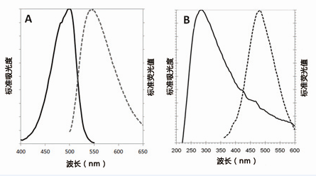
CYTO-ID@绿色检测试剂2(A组)在PBS确定的吸光度和荧光图谱(499/548nm)。
Hoechst33342(图B)在1X测定缓中液来确定的吸光度和荧光图谱(350/461nm)。

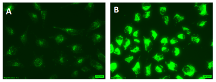
HeLa cells使用CYTO-ID@ Green Detection Reagent 2进行染色
(A)完全培养基(B)在饥饿培养基(EBSS)中添加40 uM Chloroquine培养4h。在含有Chloroquine的EBSS培养基中培养的细胞表现非常明亮的绿色荧光信号和点状结构。

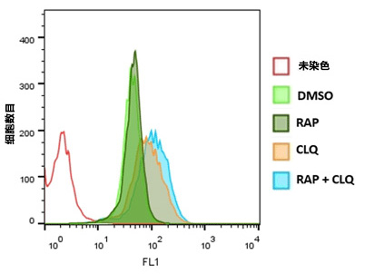
运用流式细胞术分析Jurkat细胞的自噬
通过0.5μM Rapamycin(RAP),10 uM Chloroquine(CLQ)处理或不处理Jurkat细胞,或两者一起处理20h。CYTO-ID@Green Detection Reagent2染色30min后,洗脱,用流式细胞仪分析。直方图叠加呈现结果。用RAP+CLQ处理细胞显示荧光有升高。

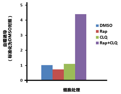
微孔板分析HepG2细胞的自噬
HepG2经过DMSO(对照),0.5uM Rapamy cin(Rap),10 uM Chloroquine(CLQ),或0.5 uM Ra p和10uMCLQ同时培养20h。细胞经过Hoechst33342染色进行细胞数目标准化。Rap和CLQ同时处理细胞显示自噬作用上升。
Product Specification
| Applications: | Flow Cytometry, Fluorescence microscopy, Fluorescent detection, HTS
|
| Application Notes: | The CYTO-ID® Autophagy detection kit 2.0 provides a rapid, specific, and quantitative approach for monitoring autophagy in live cells by fluorescence microscopy, flow cytometry, and microplate reader. |
| Quality Control: | A sample from each lot of CYTO-ID® Autophagy detection kit 2.0 is used to stain Jurkat cells and analyzed by flow cytometry, using the procedures described in the user manual. The MAF values for the samples were greater than 25. The percentage of viable cells in the control samples is > 90% and > 80% in the treated samples. |
| Quantity: | For -0200 size:
200 flow cytometry assays, 250 microscopy assays or 3 x 96-well microplate assays.
For -0050 size:
50 flow cytometry assays, 60 microscopy assays or 1 x 96-well microplate assays. |
| Use/Stability: | With proper storage, the kit components are stable for one year from date of receipt. |
| Handling: | Protect from light. Avoid freeze/thaw cycles. |
| Shipping: | Shipped on Dry Ice |
| Short Term Storage: | -20°C |
| Long Term Storage: | -80°C |
| Contents: | CYTO-ID® Green Detection Reagent 2
Hoechst 33342 Nuclear Stain
Autophagy Inducer (Rapamycin)
Chloroquine Control
10X Assay Buffer |
| Scientific Background: | Autophagy is a stress-induced protective mechanism. Less active under basal conditions, the mechanism is utilized by eukaryotic cells through lysosome-mediated bulk degradation of cellular contents when subjected to certain hostile conditions such as nutrient depletion and chemical or environmental stress. The role of increased autophagic activity in the pathology of cancer, neurodegeneration, cardiovascular disease and diabetes has become widely recognized and commonly studied. Induction of autophagic flux can be visualized by enhanced accumulation of autophagic vesicles if lysosomal function is inhibited, preventing removal of these vesicles. |
| Technical Info/Product Notes: | The CYTO-ID® Autophagy Detection kit 2.0 is a member of the CELLESTIAL® product line, reagents and assay kits comprising fluorescent molecular probes that have been extensively benchmarked for live cell analysis applications. |
| Protocol: | A detailed protocol for FC in primary BMDCs can be found on bioprotocol.org: Flow Cytometric Analysis of Autophagic Activity with CYTO-ID Staining in Primary Cells by M. Stankov, et al. |
Product Literature References
Conidiobolus coronatus induces oxidative stress and autophagy response in Galleria mellonella larvae: M. Kazek, et al.; PLoS One
15, e0228407 (2020),
Abstract;
Full TextAirway epithelial regeneration requires autophagy and glucose metabolism: K. Li, et al.; Cell Death Dis.
10, 875 (2019),
Abstract;
Full Text Aqueous extract of clove inhibits tumor growth by inducing autophagythrough AMPK/ULK pathway: C. Li, et al.; Phytother. Res.
33, 1794 (2019),
Application(s): Flow Cytometry analysis in human pancreatic and colon cancer cells (ASPC-1 and HT-29),
Abstract;
Full Text Autophagy differentially regulates macrophage lipid handling depending on the lipid substrate (oleic acid vs. acetylated-LDL) and inflammatory activation state: S. Hadadi-Bechor, et al.; Biochim. Biophys. Acta Mol. Cell Biol. Lipids
11, 158527 (2019),
Abstract;
Autophagy induction and PDGFR-β knockdown by siRNA-encapsulated nanoparticles reduce chlamydia trachomatis infection: S. Yang, et al.; Sci. Rep.
9, 1306 (2019),
Application(s): Human Vaginal Epithelial Cells (CK2/E6E7)- Autophagic Flux,
Abstract;
Full Text Decreased Autophagy Impairs Decidualization of Human Endometrial Stromal Cells: A Role for ATG Proteins in Endometrial Physiology: A.C. Mestre Citrinovitz, et al.; Int. J. Mol. Sci.
20, 3066 (2019),
Application(s): Flow Cytometry analysis in human endometrial stromal cells (t-HESC),
Abstract;
Full Text Human-Induced Pluripotent Stem Cell Model of Trastuzumab-Induced Cardiac Dysfunction in Patients With Breast Cancer: T. Kitani, et al.; Circulation
139, 2451 (2019),
Abstract;
Inhibited autophagy may contribute to heme toxicity in cardiomyoblast cells: A. Gyongyosi, et al.; Biochem. Biophys. Res. Commun.
511, 732 (2019),
Application(s): Autophagic process in H9c2 cardiomyoblast cell,
Abstract;
LKB1/p53/TIGAR/autophagy-dependent VEGF expression contributes to PM2.5-induced pulmonary inflammatory responses: H. Xu, et al.; Sci. Rep.
9, 16600 (2019),
Abstract;
Full Text miR-20a inhibits hypoxia-induced autophagy by targeting ATG5/FIP200 in colorectal cancer: J. Che, et al.; Mol. Carcinog.
58, 1234 (2019),
Application(s): Flow Cytometry and Microscopy in colon cancer cells ,
Abstract;
Zebrafish modeling of intestinal injury, bacterial exposures, and medications defines epithelial in vivo responses relevant to human inflammatory bowel disease: L.S. Chuang, et al.; Dis. Model. Mech. dmm.037432 (2019),
Application(s): Microscopy in zebrafish larvae gut,
Abstract;
Zn-doped CuO nanocomposites inhibit tumor growth by NF-κB pathwaycross-linked autophagy and apoptosis: H. Xu, et al.; Nanomedicine (Lond.)
14, 131 (2019),
Application(s): Flow Cytometry and Microscopy in cancer cells (Panc28 and HepG2),
Abstract;
Genistein promotes ionizing radiation-induced cell death by reducing cytoplasmic Bcl-xL levels in non-small cell lung cancer: Z. Zhang, et al.; Sci. Rep.
8, 328 (2018),
Abstract;
Full Text Knockout of ATG5 leads to malignant cell transformation and resistance to Src family kinase inhibitor PP2: S.H. Hwang, et al.; J. Cell. Physiol.
233, 506 (2018),
Abstract;
Maslinic acid induces autophagy by down-regulating HSPA8 in pancreatic cancer cells: Y. Tian, et al.; Phytother. Res.
32, 1320 (2018),
Abstract;
Metabolite of ellagitannins, urolithin A induces autophagy and inhibits metastasis in human sw620 colorectal cancer cells: W. Zhao, et al.; Mol. Carcinog.
57, 193 (2018),
Abstract;
Silver nanoparticles induce lysosomal-autophagic defects and decreased expression of transcription factor EB in A549 human lung adenocarcinoma cells: T. Miyayama, et al.; Toxicol. In Vitro.
46, 148 (2018),
Application(s): Confocal microscopy with A549 cells,
Abstract;
The Oxysterol 7-Ketocholesterol Reduces Zika Virus Titers in Vero Cells and Human Neurons: K.A. Willard, et al.; Viruses
11, 20 (2018),
Application(s): Flow Cytometry in Vero cells,
Abstract;
Full Text Alisertib promotes apoptosis and autophagy in melanoma through p38 MAPK-mediated aurora a signaling: Y. Shang, et al.; Oncotarget.
8, 107076 (2017),
Abstract;
Full Text Humic acid inhibits HBV-induced autophagosome formation and induces apoptosis in HBV-transfected Hep G2 cells: K. Pant, et al.; Sci. Rep.
6, 34496 (2016),
Application(s): Autophagosome formation assay, HepG2 cells,
Abstract;
Full Text Inhibition of autophagy with chloroquine potentiates carfilzomib-induced apoptosis in myeloma cells in vitro and in vivo: V. Jarauta, et al.; Cancer Lett.
382, 1 (2016),
Abstract;
General Literature References
A live-cell fluorescence microplate assay suitable for monitoring vacuolation arising from drug or toxic agent treatment: J. Coleman, et al.; J. Biomol. Screen.
15, 398 (2010),
Abstract;
Methods in mammalian autophagy research: N. Mizushima, et al.; Cell
140, 313 (2010),
Abstract;
Assays to Assess Autophagy Induction and Fusion of Autophagic Vacuoles with a Degradative Compartment, Using Monodansylcadaverine (MDC) and DQ-BSA: C.L. Vazquez & M.I. Colombo; Methods Enzymol.
452, 85 (2009),
Abstract;
Desmethylclomipramine induces the accumulation of autophagy markers by blocking autophagic flux: M. Rossi, et al.; J. Cell Sci.
122, 3330 (2009),
Abstract;
Guidelines for the use and interpretation of assays for monitoring autophagy in higher eukaryotes: D.J. Klionsky, et al.; Autophagy
4, 151 (2008),
Abstract;


